

Case Study

This case involves a 52-year-old patient with long-segment, non-dysplastic Barrett’s esophagus (NDBE) and low-grade esophagitis. His clinical and histologic characteristics suggested the patient was low-risk for progression to high-grade dysplasia (HGD) or esophageal adenocarcinoma (EAC), but after a concerning increase in his BE segment length during a repeat endoscopy, Dr. Muthusamy used TissueCypher to evaluate the patient’s personalized risk of progression.  

“This is a patient that we were able to intervene in early before he developed any dysplasia.”  

See more details below about how Dr. Muthusamy navigated this case.

Case details

  • No relevant family history
  • No obesity
  • Chronic GERD, on PPIs
  • Non-smoker

EGD performed one year prior found:  

  • Hiatal hernia
  • 5 cm esophageal stricture with associated esophagitis in the distal esophagus

Case Study

This case involves a 52-year-old patient with long-segment, non-dysplastic Barrett’s esophagus (NDBE) and low-grade esophagitis. His clinical and histologic characteristics suggested the patient was low-risk for progression to high-grade dysplasia (HGD) or esophageal adenocarcinoma (EAC), but after a concerning increase in his BE segment length during a repeat endoscopy, Dr. Muthusamy used TissueCypher to evaluate the patient’s personalized risk of progression.  

“This is a patient that we were able to intervene in early before he developed any dysplasia.”  

See more details below about how Dr. Muthusamy navigated this case.

Case details

  • No relevant family history
  • No obesity
  • Chronic GERD, on PPIs
  • Non-smoker

EGD performed one year prior found:  

  • Hiatal hernia
  • 5 cm esophageal stricture with associated esophagitis in the distal esophagus

Case Study

This case involves a 52-year-old patient with long-segment, non-dysplastic Barrett’s esophagus (NDBE) and low-grade esophagitis. His clinical and histologic characteristics suggested the patient was low-risk for progression to high-grade dysplasia (HGD) or esophageal adenocarcinoma (EAC), but after a concerning increase in his BE segment length during a repeat endoscopy, Dr. Muthusamy used TissueCypher to evaluate the patient’s personalized risk of progression.  

“This is a patient that we were able to intervene in early before he developed any dysplasia.”  

See more details below about how Dr. Muthusamy navigated this case.

Case details

  • No relevant family history
  • No obesity
  • Chronic GERD, on PPIs
  • Non-smoker

EGD performed one year prior found:  

  • Hiatal hernia
  • 5 cm esophageal stricture with associated esophagitis in the distal esophagus
Blue gradient background with vertical rounded lines in varying shades of blue scattered across the right side.
Dr. Raman Muthusamy: Case 1
Bearded man with glasses in blue blazer and shirt speaking in front of a blurred abstract painting.

Visually low-risk, biologically high-risk

This case involves a 52-year-old patient with long-segment, non-dysplastic Barrett’s esophagus (NDBE) and low-grade esophagitis. His clinical and histologic characteristics suggested the patient was low-risk for progression to high-grade dysplasia (HGD) or esophageal adenocarcinoma (EAC), but after a concerning increase in his BE segment length during a repeat endoscopy, Dr. Muthusamy used TissueCypher to evaluate the patient’s personalized risk of progression.  

“This is a patient that we were able to intervene in early before he developed any dysplasia.”  

See more details below about how Dr. Muthusamy navigated this case.

Clinical risk factors

A 52-year-old, white, male patient in very good physical condition had a history of chronic GERD and long-term PPI use with limited symptom control. He was also diagnosed with a hiatal hernia and long-segment NDBE.

RELEVANT MEDICAL HISTORY
  • No relevant family history
  • No obesity
  • Chronic GERD, on PPIs
  • Non-smoker

Endoscopic findings

The initial endoscopy confirmed long-segment, NDBE. A follow-up endoscopy found an increase in segment length.

Lower esophagus:

  • Erosive esophagitis (Los Angeles Grade B)  
  • 3 cm hiatal hernia (Hill Grade 4)
  • Barrett’s segment length: C6M6  
    (Follow-up endoscopy found segment to be C7M8)
  • Pathology returned non-dysplastic  

Traditional Management:

  • Treatment recommendation: 3-year surveillance

TissueCypher results

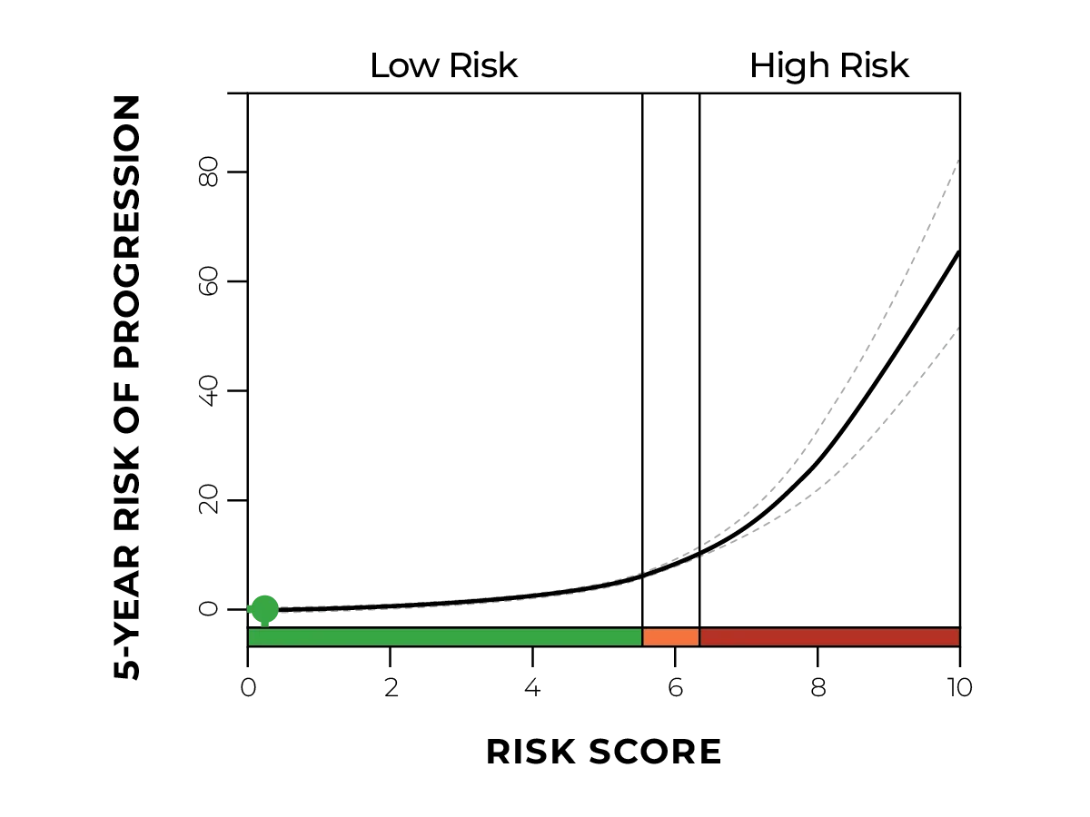
Despite the patient’s relatively young age and good physical shape, the TissueCypher test returned a high-risk score of 7.3 with a 5-year risk of progression to HGD/EAC of 19%. Dr. Muthusamy confidently escalated the patient to ablation to avoid disease progression.  

  • Risk class: High
  • Risk score: 7.3
  • 5-year probability of progression: 19%

 TissueCypher-guided management

  • Escalate to radiofrequency ablation (RFA)
  • Completion of C-TIF for reflux control

"We were planning on probably a three-year surveillance, but his TissueCypher score came back as actually high risk. That fact, coupled with his concerns about his difficult to control reflux and his large hiatal hernia spurred this patient to undergo action.”

Raman Muthusamy, MD