











Man and woman in life jackets paddling a green canoe on a calm lake with forested hills behind.
How it works

Informs risk appropriate management

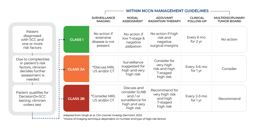
The integrated DecisionDx-SCC test result combines the 40-gene expression profile with clinicopathologic risk factors and optimized nested predictive models to deliver refined risk-assessment across 4 result classes.

  1. Test ordered

    Biopsy from a patient with high-risk cutaneous SCC

  2. Optimized nested predictive models

    The i40-GEP

    Gene expression algorithm

    The molecular signature is based on 40 genes

    Clinicopathologic factors

    Immunosuppression, tumor diameter, differentiation, PNI, and anatomic site

    Integrated into a single, validated risk classification.

  3. Four risk classes

    Class 1A
    Low risk
    Class 1B
    Moderate risk
    Class 2A
    High risk
    Class 2B
    Highest risk
  1. Test ordered

    Biopsy from a patient with high-risk cutaneous SCC

  2. Optimized nested predictive models

    The i40-GEP

    Gene expression algorithm

    The molecular signature is based on 40 genes

    Clinicopathologic factors

    Immunosuppression, tumor diameter, differentiation, PNI, and anatomic site

    Integrated into a single, validated risk classification.

  3. Four risk classes

    Class 1A
    Low risk
    Class 1B
    Moderate risk
    Class 2A
    High risk
    Class 2B
    Highest risk

DecisionDx-SCC testing process

DecisionDx-SCC is performed in Castle Biosciences’ CLIA certified, CAP-accredited and NY state approved laboratory using formalin-fixed, paraffin embedded (FFPE) primary tumor tissue from either a biopsy or excision.

Green microscope icon on white pixelated background.

Tissue biopsy submitted

Tissue blocks or slides from the primary tumor tissue biopsies are sent to our laboratory for testing.

Pixelated green and white circular design with diagonal stripes on black background.

RNA/DNA preparation

Once tissue is received in our laboratory, RNA is isolated from the tissue and converted to cDNA. The cDNA for each of the 40 genes is amplified and the gene signature recorded.

Pixel art key with a white head and green shading on the shaft.

Signature interpretation

By combining the 40-gene expression profile with clinicopathologic risk factors and optimized nested predictive models a class result is provided.

Green abstract geometric shapes forming a stylized letter S on a white background.

DecisionDx-SCC reporting

The integrated DecisionDx-SCC report provides a class result based on a patient’s risk for metastasis and local recurrence:

  • Class 1: Low risk

  • Class 1B: Moderate risk

  • Class 2A: High risk

  • Class 2B: Highest risk

Download Guide

Provides precise, personalized risk to inform patient management & treatment guidance ​

40-GEP

PNI, poor differentiation, immunosuppression, tumor diameter and tumor location

i40-GEP result
(MFS on validation cohort)

Clinical utility
Based on metastatic risk

  1. Class 1A

    Low risk

    MFS 97.3% | LRFS 96.8%

    ART benefit

    Low likelihood

    0–5% metastatic risk

    • Safe to defer imaging
    • Safe to defer ART
  2. Class 1B

    Moderate risk

    MFS 94.6% | LRFS 90.0%

    ART benefit

    Low likelihood

    5–10% metastatic risk

    • Consider increased surveillance
    • Consider imaging
    • Safe to defer ART
  3. Class 2A

    High risk

    MFS 77.9% | LRFS 83.1%

    ART benefit

    Low likelihood

    >20% metastatic risk

    • Recommend increased surveillance
    • Recommend imaging
    • Recommend multidisciplinary consult
    • Unlikely benefit from ART
  4. Class 2B

    Highest risk

    MFS 66.2% | LRFS 82.1%

    ART benefit

    High likelihood

    >20% metastatic risk

    • Recommend increased surveillance
    • Recommend imaging
    • Recommend multidisciplinary consult
    • Likely benefit from ART

40-GEP

PNI, poor differentiation, immunosuppression, tumor diameter and tumor location

i40-GEP result
(MFS on validation cohort)

Clinical utility
Based on metastatic risk

  1. Class 1A

    Low risk

    MFS 97.3% | LRFS 96.8%

    ART benefit

    Low likelihood

    0–5% metastatic risk

    • Safe to defer imaging
    • Safe to defer ART
  2. Class 1B

    Moderate risk

    MFS 94.6% | LRFS 90.0%

    ART benefit

    Low likelihood

    5–10% metastatic risk

    • Consider increased surveillance
    • Consider imaging
    • Safe to defer ART
  3. Class 2A

    High risk

    MFS 77.9% | LRFS 83.1%

    ART benefit

    Low likelihood

    >20% metastatic risk

    • Recommend increased surveillance
    • Recommend imaging
    • Recommend multidisciplinary consult
    • Unlikely benefit from ART
  4. Class 2B

    Highest risk

    MFS 66.2% | LRFS 82.1%

    ART benefit

    High likelihood

    >20% metastatic risk

    • Recommend increased surveillance
    • Recommend imaging
    • Recommend multidisciplinary consult
    • Likely benefit from ART

40-GEP

PNI, poor differentiation, immunosuppression, tumor diameter and tumor location

i40-GEP result
(MFS on validation cohort)

Clinical utility
Based on metastatic risk

  1. Class 1A

    Low risk

    MFS 97.3% | LRFS 96.8%

    ART benefit

    Low likelihood

    0–5% metastatic risk

    • Safe to defer imaging
    • Safe to defer ART
  2. Class 1B

    Moderate risk

    MFS 94.6% | LRFS 90.0%

    ART benefit

    Low likelihood

    5–10% metastatic risk

    • Consider increased surveillance
    • Consider imaging
    • Safe to defer ART
  3. Class 2A

    High risk

    MFS 77.9% | LRFS 83.1%

    ART benefit

    Low likelihood

    >20% metastatic risk

    • Recommend increased surveillance
    • Recommend imaging
    • Recommend multidisciplinary consult
    • Unlikely benefit from ART
  4. Class 2B

    Highest risk

    MFS 66.2% | LRFS 82.1%

    ART benefit

    High likelihood

    >20% metastatic risk

    • Recommend increased surveillance
    • Recommend imaging
    • Recommend multidisciplinary consult
    • Likely benefit from ART

References: Ratner et al, AAD Innovation Academy 2026, Shah M, et al. J of Skin. 2025, Patel VA, et al. Cancer Med. 2022, Rentroia-Pacheco B, et al.eClinicalMedicine. 2023, Gupta N, et al. J Am Acad Dermatol. 2022, Wang DM, et al. JAMA Dermatol. 2025. *Report distribution experienced in validation.

Get started with DecisionDx-SCC

NEXT UP

Scientific Evidence